CASE REPORT
https://doi.org/10.47811/bhj.215
Isolated hoarseness revealing an occult extensive aortic dissection - A rare case of Ortner Syndrome
1Department of ENT and Head Neck Surgery, Jigme Dorji Wangchuck National Referral Hospital, Thimphu, Bhutan
2Department of Medicine, Jigme Dorji Wangchuck National Referral Hospital, Thimphu, Bhutan
Corresponding author
Dr. Sonam Jamtsho
ABSTRACT
Hoarseness of voice due to vocal cord palsy may indicate an underlying sinister pathology. However, vocal cord paralysis as the sole manifestation of aortic dissection and aneurysm is rare. We report the case of a 63-year-old man with diabetes mellitus, hypertension and a 40-year smoking history, who presented with hoarseness to the ENT Outpatient Department of Jigme Dorji Wangchuck National Referral Hospital. Evaluation revealed left vocal cord palsy, and a chest X-ray showing mediastinal widening prompted further imaging. Contrast-enhanced computed tomography revealed extensive aortic dissection. A multidisciplinary discussion involving cardiology, cardiothoracic surgery, ENT and the patient's family led to a decision of conservative management with strict blood pressure control and smoking cessation.
INTRODUCTION
Hoarseness of voice is a common presenting complaint in otolaryngology. It results from incomplete closure of vocal cords during phonation and may be caused by conditions such as acute or chronic laryngitis, gastroesophageal reflux disease, vocal cord tumours or laryngeal nerve paralysis due to various causes. Nobert Ortner first described three cases of left vocal cord palsy caused by compression of the left Recurrent Laryngeal Nerve (RLN) by an enlarged left atrium secondary to severe mitral stenosis1. Now known as Ortner Syndrome or cardiovocal syndrome, it is associated with a wide range of cardiovascular disorders like congenital heart disease, aortic aneurysms, aortic dissection, left atrial myxoma and severe pulmonary hypertension2. Aortic dissection typically presents with sudden shearing chest, back or abdominal pain, and may also present as syncope, stroke, or end-organ ischemia3. However, aortic dissection presenting as isolated hoarseness due to RLN paralysis is very rare4-7. We report a case of Ortner Syndrome in which an isolated hoarseness was the sole presenting feature of an extensive occult aortic dissection.
PATIENT INFORMATION
A 63-year-old man with a history of type 2 diabetes mellitus and hypertension presented to the Department of Otorhinolaryngology with progressive hoarseness of voice for two months. The onset was insidious, beginning with mild throat discomfort, and gradually progressing to persistent dysphonia with a breathy vocal quality. He denied odynophagia, dysphagia, fever, cough, haemoptysis, dyspnoea, or chest pain. His hypertension was well controlled with antihypertensive medication for 10 years, and his diabetes was managed with oral hypoglycaemic agents. He was a chronic smoker with a 40-year smoking history.
CLINICAL FINDINGS
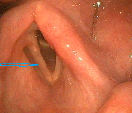
On examination, the patient was clinically stable with an Eastern Cooperative Oncology Group (ECOG) performance status of 0. Examination of the nasal cavity, oral cavity, head and neck, and other systems were unremarkable. Fiberoptic laryngoscopy revealed left vocal cord paralysis with mild atrophy of the left vocal fold and a significant phonatory gap resulting in a breathy voice. The remaining laryngeal and hypopharyngeal structures appeared normal as shown in figure 1.
DIAGNOSTIC ASSESSMENT
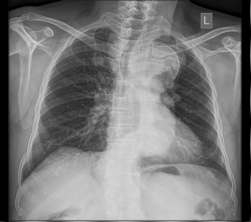
Given his chronic smoking history and to evaluate for possible intrathoracic pathology, a chest radiograph was obtained. It demonstrated a widened mediastinum, a calcified aortic knuckle, dilated tortuous descending thoracic aorta, and rightward tracheal deviation as shown in Figure 2.
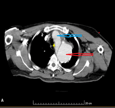
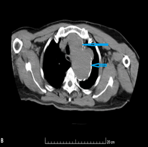
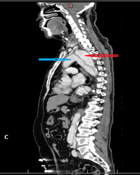
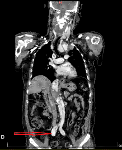
Transthoracic echocardiography demonstrated dilatation of the aortic arch (4.5 cm) and descending thoracic aorta (5.0 cm), with preserved left ventricular systolic function and an ejection fraction of 65%. Contrast-enhanced computed tomography (CECT) of the neck and chest revealed an extensive aortic dissection originating in the aortic arch just distal to the origin of the left subclavian artery and extending to the level of the iliac artery, consistent with a Stanford type B aortic dissection. The thoracic aorta was markedly dilated, measuring 6.4 cm in maximum diameter. A large false lumen measuring 4.96 cm compressed the true lumen as shown in figures 3A to 3D. The right renal artery and common iliac artery were noted to arise from the false lumen.
THERAPEUTIC INTERVENTION
The patient was managed by a multidisciplinary team comprising ENT, cardiology and cardiothoracic surgical specialists. Intensive medical therapy was initiated for optimal blood pressure control. Given the extensive aortic dissection and a heavily calcified aorta, no active surgical intervention was instituted due to the high procedural risk and limited expected benefit. The patient and his family members were counselled regarding the condition and advised to stop smoking, adhere to antihypertensive therapy, and avoid heavy lifting and strenuous activity. His dysphonia was managed conservatively as there was no evidence of aspiration of food or drinks.
FOLLOW - UP AND OUTCOMES
At the three-month follow-up, the patient reported mild subjective improvement in his voice. He denied aspiration of food or liquids and had no breathing difficulties. Fiber optic laryngoscopy showed left vocal cord atrophy with good compensation by the right vocal cord. He reported no limb or abdominal pain. He had continued regular follow-up for hypertension and diabetes, with blood pressure and blood sugar levels remaining within the normal range.
DISCUSSION
Aortic dissection is a grave emergency with a significant mortality of 18.8% for Stanford type A and 13.3% for Stanford type B dissections8. The follow up mortality rates of the type B dissections are relatively high approaching about 1 in 4 patients in 3 years9.
RLN paralysis may arise from a wide variety of causes, including malignancy, trauma, iatrogenic injury and idiopathic conditions. Lung cancers (42%) followed by iatrogenic injury during the surgery (42%) attributes to the majority of cases1. Both RLNs arise from the vagus nerve and innervate the intrinsic muscles of the larynx. The right RLN loops around the right subclavian artery, while the left RLN descends into the thorax and loops beneath the aortic arch before ascending in the tracheoesophageal loop. The left RLN is particularly vulnerable to injury owing to its longer and more complex intrathoracic course. Furthermore, its close anatomical relationship with major cardiovascular structures makes it susceptible to compression or stretching from enlarged or distorted cardiovascular structures, forming the basis of cardio vocal syndrome.
While the majority of patients with aortic dissection present with pain, up to 10% have a silent or painless presentation6,10. In rare cases, hoarseness of voice may be the only presenting symptom11-12. Our patient had no history of shearing chest pain or discomfort and presented solely with hoarseness of voice. This occurs due to the compression of the RLN as it hooks around the aortic arch, due to the external compression from an expanding hematoma or an expanding dissecting aneurysm4. Although initially described in association with mitral stenosis, Ortner Syndrome has since been reported in a wide range of cardiovascular conditions like pulmonary hypertension, silent aortic dissection and pulmonary diseases like chronic obstructive pulmonary disease13-15.
Hoarseness of voice is commonly caused by benign conditions such as vocal overuse, laryngitis, gastroesophageal reflux disease or vocal cord lesions. Vocal cord paralysis must be confirmed by an ENT surgeon after a thorough ENT examination. When routine evaluation fails to identify an obvious cause, an underlying cardiovascular etiology should be considered. In rare instances, hoarseness of voice may be the only clinical manifestation of serious underlying cardiovascular disease, as demonstrated in our patient with extensive aortic dissection.
Surgical intervention is indicated for Stanford type A dissections and for complicated type B dissections16. Surgical treatment of aortic dissection carries a significant risk, with reported mortality rates of up to 21.6% with lower volume centers having double the rates of risk-adjusted mortality compared to those performed by high volume providers17. Management of vocal cord paralysis depends on the severity of symptoms, impact on quality of life and the risk of aspiration. If indicated, medialization of the vocal cord is the main surgical treatment option considered1,9. In patients without aspiration or recurrent lung infections, conservative management with regular reassessment of laryngeal function is appropriate2. In our patient, vocal cord paralysis resulted only in hoarseness of voice. He was able to communicate and had preserved laryngeal function. Therefore, he was managed conservatively with scheduled follow-up. At his three-month follow-up, he reported mild improvement in voice, with laryngoscopy demonstrating good compensatory function of the contralateral vocal cord.
CONCLUSION
This case highlights the importance of maintaining a high index of suspicion for serious underlying cardiovascular pathology in patients presenting with sudden onset hoarseness of voice. Although rare, silent aortic dissection can manifest solely as hoarseness of voice. Early recognition, appropriate imaging and timely multidisciplinary evaluation involving otolaryngology and cardiology are crucial, as isolated hoarseness may be the only manifestation of serious cardiovascular disease.
INFORMED CONSENT
Written informed consent was obtained from the patient for the use of clinical information and images. All personal identifiers have been removed to ensure patient confidentiality.
REFERENCES
- Kyaga DH, de Souza GR, de Almeida JHRR, Nobeschi L, Burihan MC. A Systematic Review of Ortner's Syndrome. J US-China Med Sci. 2018;15(1):10-15. [Full Text] [DOI
- Verma S, Talwar A, Talwar A, Khan S, Krishnasastry KV, Talwar A. Ortner's syndrome: A systematic review of presentation, diagnosis and management. Intractable Rare Dis Res. 2023;12(3):141-7. [PubMed] [Full Text] [DOI
- Attia RQ, Jassar AS. Acute Aortic Dissection. In: Near Misses in Cardiac Surgery. Cham: Springer International Publishing; 2022. p. 191- 4.
- Semionov A, Kosiuk J. Ortner syndrome secondary to aortic aneurysm. Radiol Case Rep. 2016;12(1):29-30. [PubMed] [Full Text] [DOI
- Passi P, Singh S, Passi B. Large fusiform aortic aneurysm with mural thrombus - a rare cause of hoarseness of voice. Indian J Med Spec. 2017;8(4):206-8. [DOI
- Rizvi M, Singh R, Jain A, Sarkar A. Asymptomatic aortic aneurysm causing right vocal cord palsy and hoarseness: A rare presentation. Anesth Essays Res. 2014;8(3):397. [PubMed] [Full Text] [DOI
- Bayones M, Ruiz-Rodriguez J, Sierra C. Aortic aneurysm as a cause of dysphonia in a patient who smokes. P R Health Sci J. 2020;39(2):229-31. [PubMed] [Full Text
- Tien M, Ku A, Martinez-Acero N, Zvara J, Sun EC, Cheung AT. The Penn classification predicts hospital mortality in acute Stanford type A and type B aortic dissections. J Cardiothorac Vasc Anesth. 2020;34(4):867-73. [PubMed] [Full Text] [DOI
- Tsai TT, Fattori R, Trimarchi S, Isselbacher E, Myrmel T, Evangelista A, et al. Long-term survival in patients presenting with type B acute aortic dissection: insights from the International Registry of Acute Aortic Dissection. Circulation. 2006;114(21):2226-31. [PubMed] [Full Text] [DOI
- Gnagi SH, Howard BE, Hoxworth JM, Lott DG. Acute Contained Ruptured Aortic Aneurysm Presenting as Left Vocal Fold Immobility. Case Rep Otolaryngol. 2015;2015:219090. [PubMed] [Full Text] [DOI
- Khan IA, Wattanasauwan N, Ansari AW. Painless aortic dissection presenting as hoarseness of voice: Cardiovocal syndrome: Ortner's syndrome. Am J Emerg Med. 1999;17(4):361-3. [PubMed] [DOI
- Chen HC, Lin CJ, Tzeng YS, Tsai CS, Wang CH. Hoarseness as an unusual initial presentation of aortic dissection. Eur Arch Otorhinolaryngol. 2005;262(3):189-91. [PubMed] [DOI
- Shankar O, Lohiya BV. Cardiovocal syndrome - A rare presentation of primary pulmonary hypertension. Indian Heart J. 2014;66(3):375-7. [PubMed] [Full Text] [DOI
- Eccles SR, Banks J, Kumar P. Ascending aortic aneurysm causing hoarse voice: a variant of Ortner's syndrome. BMJ Case Rep. 2012;3-5. [PubMed] [Full Text] [DOI
- Rajamani SK, Iyer RS, Karodpati NS. Etiopathogenesis of Cardiovocal Syndrome (Ortner's Syndrome): A Systematic Review of the Literature. Indian J Clin Cardiol. 2023;4(2):101-9. [Full Text] [DOI
- Sawa A, Shirokawa T, Kobayashi H, Satoh H. Ortner's syndrome in a patient with COPD. Intern Med. 2012;51(15):2059. [PubMed] [Full Text] [DOI
- Carrel T, Sundt TM, von Kodolitsch Y, Czerny M. Acute aortic dissection. The Lancet. 2023;401(10378):773-88. [PubMed] [DOI
- Chikwe J, Cavallaro P, Itagaki S, Seigerman M, Diluozzo G, Adams DH. National outcomes in acute aortic dissection: influence of surgeon and institutional volume on operative mortality. Ann Thorac Surg. 2013;95(5):1563-9. [PubMed] [Full Text] [DOI
|
AUTHORS CONTRIBUTION Following authors have made substantial contributions to the manuscript as under: SJ: Conceptualization, data collection, manuscript writing. SD: Data collection, manuscript writing. Authors agree to be accountable for all respects of the work in ensuring that questions related to the accuracy and integrity of any part of the work are appropriately investigated and resolved. |
|
CONFLICT OF INTEREST None GRANT SUPPORT AND FINANCIAL DISCLOSURE None |